Ultra Wide-field Imagaing
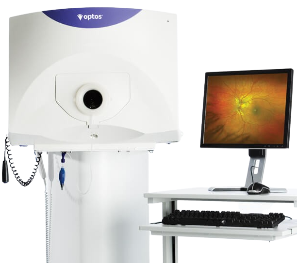
Optos Retinal Imaging
Your eyes are the window to your overall health.
As part of your annual eye exam our doctors exam not only check your prescription, but more importantly the health of your eyes. For many of us growing up, dilation was the only option for doctors to see into our eyes causing temporary light sensitivity and blurry vision.
We offer the Optos ultra-wide digital retinal imaging system as a clinical alternative to dilation in many cases. This technology allows us to have year over year records of your eye health for both monitoring and patient education.
Optos can capture up to 80% of your retina and far periphery with its advanced 200° panoramic imaging capabilities in a single shot. This aids in the earliest detection, treatment, and management of diseases such as Diabetes, Age Related Macular Degeneration (AMD), and other conditions that can affect both your eyes and your overall health.
Adding this latest technology to your appointment will help you and your eye doctor make educated decisions about your overall care.
Does Optos Imaging Replace Dilation?
While Optos imaging is an amazing technology for capturing the retina much more effectively than traditional devices, there are some situations where you and your doctor’s clinical evaluations may benefit from dilation as well. For far peripheral retinal views close to the ora serrata, dilation and indirect ophthalmoscopy will allow your eye doctor to see further out when needed.
What is a retina?
The retina is a thin layer of tissue that lines the back of the eye on the inside. It is located near the optic nerve. The purpose of the retina is to receive light that your eye has focused and convert into a message that is sent to the brain. This message will then be interpreted as a visual image or object.
Early and regular monitoring of the retina can help your doctor to signs of impairment or even retinal detachments.
Retinal Detachment
A retinal detachment is when the retina separates from the back of the eye. Symptoms may be sudden flashes of light, intense floaters, and blind spots in the vision or total loss of vision.*
Patients with high myopia are more likely to develop potentially sight threatening Retinal Detachments. Being able to image the peripheral retina is key to detecting retinal holes and tears which can lead to Retinal Detachments.
A dilated examination, with the addition of a photographic images with Optos, will reveal a more accurate portrayal of the immediate condition and potentially urgent referral to a retinal specialist for it’s repair and management.
Retinal Tear+ Retinal Detachment

*Please contact the office immediately if you are experiencing any of these symptoms.
What are some of the overall health conditions that may be detected with the Optos?
Diabetes
Our clinic monitors diabetic patients on a 6 month rotation to partner in the successful management of your diabetes as well as to catch any early signs of Diabetic Retinopathy.
Diabetic Retinopathy is a complication from uncontrolled Diabetes.
Uncontrolled Diabetes can damage tiny blood vessels within the retina and is one of the leading causes of blindness. Early stages of this condition may not be apparent to someone with diabetes and may threaten to damage your eyesight when going unmanaged.
Utilizing the OptosPlus imaging and capturing a real-time photograph is critical for our doctors to help in your health care management. In addition, it creates a visual tool for you to see exactly what your doctor sees to enrich your understanding and convey the urgency of the condition.
Age-Related Macular Degeneration
Age-related Macular Degeneration is the leading cause of profound vision loss in people over age 60. It occurs when a small “bull’s-eye” in the retina, known as the macula, deteriorates. Because the disease develops as a person ages, it is referred to as age-related macular degeneration (AMD).
Optomap Color View of the Eye
Optomap Red-Free View of the Eye

Retinal Tear+ Retinal Detachment

Optomap Red-Free View of the Eye
High Tech Eyecare
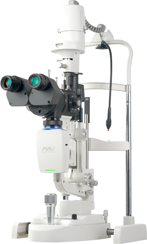
Firefly Slit LaMp
Start with an Exceptional Digital Slit Lamp
The Firefly’s Wide Dynamic Range (WDR) camera crisply delineates the structural details of your cornea, lens, iris, and other eye anatomy by using wavelengths similar to natural light.
This allows our doctors to not only have the best view during your exam, but also allows us to record high-resolution imagery in real time to help educate you as a partner in your overall health.
Optical Resolution up to 200 lp/mm
The optics in the new Firefly leap over conventional slit lamps and even other high tech slit lamps on the field today. Couple this with the high-resolution and 5 Mega Pixels video output, our doctors won’t be missing anything!
Efficiency is Automatic
Firefly was designed for the anterior segment aka the front of your eye! This allows us to run state of the art dry eye diagnostics, perform meibomian gland observation with the ability to share what we see, and enhances our Ortho-K success rates in Myopia Management.
The Firefly digital slit lamp is also an efficiency specialist with time-saving features, including:
- Automatic exposure to set your doctor up with the best view quicker!
- Auto white balance, with an adjustable depth of field and dynamic range to create vivid images for diagnosis and to share with you!
- No parameter settings required to ensure more time is spent on your care and not setting up devices.
The built-in infrared lighting system captures a larger scope in meibomian glands.
With Firefly’s clean LED light, super yellow filter and cobalt blue light, the sodium fluorescein imaging simply pops, allowing for easily viewable tear-film and corneal disease observation.
The Dry Eye Diagnostics module enhances your dry-eye care!
Firefly’s Dry Eye Module gives our doctors a strong assist to diagnose if/when you have dry eye, to identify its causes, and develop quantifiable treatment plans for you and your loved ones.


The Next Level of Contact Lens Fitting
The Orthokeratology Module improves the efficiency and accuracy of lens fitting, and a smoother experience for you and reduces the number of times you have to come in for fine tuning.
We can record high-resolution fluorescein images of your lens fitting, and real-time video without a recording limit. The module allows us to view multiple lens fit images and to show you the benefits of each!

Tagline Here
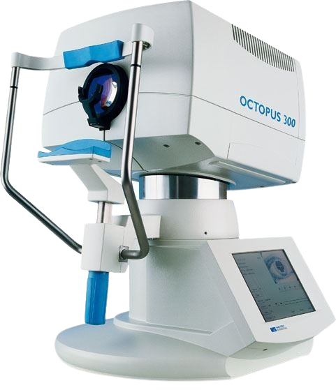
Octopus 600 Visual Field
Octopus 600 is a computer perimeter that analyzes contrast sensitivity also referred to as your visual field. Your visual field is how wide of an area your eye can see when you focus on a central point. Visual field testing is one way your doctor can measure how much vision you have in either eye, and how much vision loss may have occurred over time.
It is also used to help your doctor detect the initial stage of glaucoma development. Pathological disorders can be identified even when other parameters are within the normal range. The innovative technology of the Octopus 600 enables your technicians to capture standard diagnostic results in only 2-4 minutes.
The Octopus 600 combines the Pulsar method for early glaucoma detection and standard white-on-white perimetry for long-term follow-up in one compact and standalone device. It is the first visual field analyzer that performs standard white-on-white perimetry on an integrated flicker-free screen, using TFT and LED technology allowing your doctor to have more data points to partner in your long term vision care.
Relief is in sight
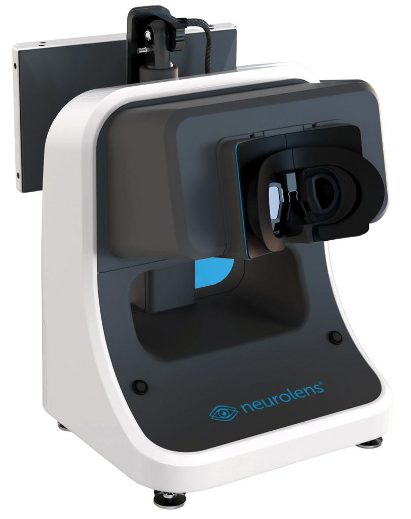
Neurolens nMD2
The Neurolens Measurement Device Gen 2 (NMD2) is an objective, accurate, and repeatable way to measure eye alignment prevalent in many patients, but often foes untreated. The industry leading eye tracking system allows the NMD2 to identify eye misalignment as small as 0.01 prism diopters, acquiring over 10,000 data points per patient in cross reference to an algorithm of thousands of similar patients.
These results allow for a customized contoured prism design placing more prism where it is needed most vs standard prism, thus correcting eye misalignment naturally and providing comfortable vision for the patients.
With the digital world demands increase; our vision demands must evolve beyond simply 20/20 when look far or near. At least 2 out of 3 people experience the symptoms of eye misalignment, and that number grows as we shift to remote working, remote learning, as well as more recreational time spent on digital devices.
Even small misalignments can cause painful symptoms, and even small prism corrections can provide dramatic relief for:
- Headaches
- Neck Pain
- Eye Strain
- Eye Fatigue
- Dry Eye Sensation
- Motion Sickness
Average screen time has increased to 13+ hours per day since March 2020, and this acceleration is already having a profound impact. Based on data from over 160,000 patients, 82% of people experience the symptoms of eye misalignment at least some of the time.
Stay Tuned!
Tagline Here
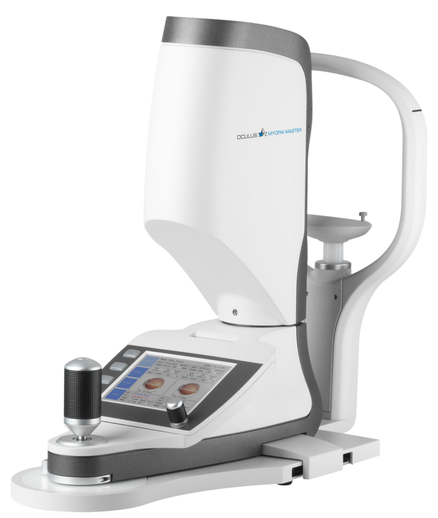
Myopia Master
Lorem ipsum dolor sit amet, consectetuer adipiscing elit. Aenean commodo ligula eget dolor. Aenean massa. Cum sociis natoque penatibus et magnis dis parturient montes, nascetur ridiculus mus. Donec quam felis, ultricies nec, pellentesque eu, pretium quis, sem. Nulla consequat massa quis enim.
Curabitur ullamcorper ultricies nisi. Nam eget dui. Etiam rhoncus. Maecenas tempus, tellus eget condimentum rhoncus, sem quam semper libero, sit amet adipiscing sem neque sed ipsum.
Stay Tuned!
Tagline Here
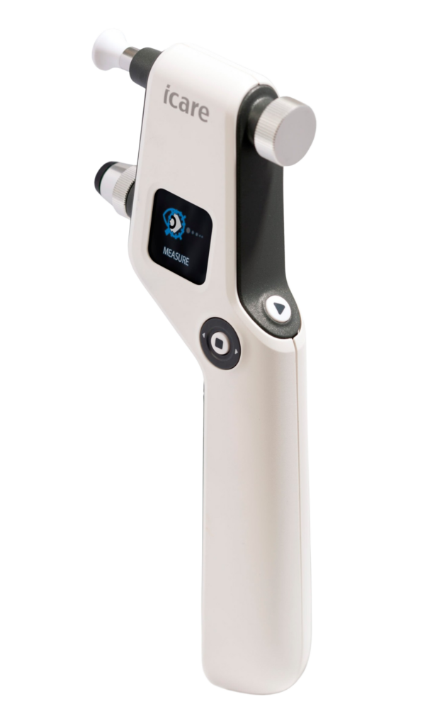
iCARE
Say goodbye to the air puff!
Stay Tuned.
Tagline Here
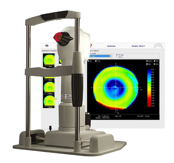
Medmont topography
More to come!
Stay tuned!


